01
Overview
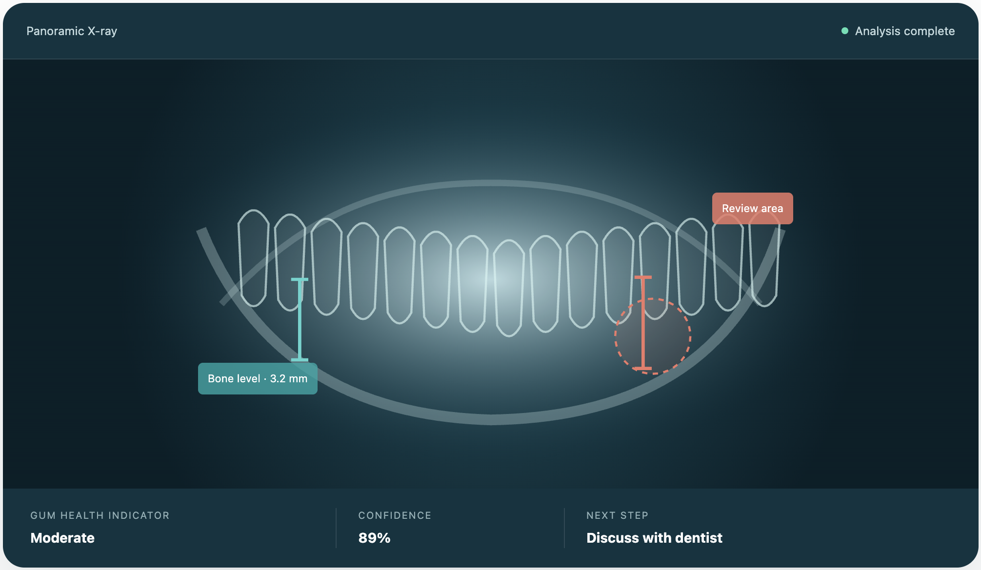
Perio ai reads a standard 2D dental panoramic X-ray and generates a plain-language gum health report with visual overlays highlighting the areas of concern. The goal is to bridge clinical expertise and patient understanding — giving dentists a consistent, explainable second read, and giving patients a report they can actually understand well enough to act on, instead of a raw X-ray and a rushed verbal explanation.

Disclaimer shown alongside every result: "AI-assisted insights for education and communication — not a replacement for professional diagnosis or treatment decisions."
02
Customer challenge
Clinical findings are clear to dentists. They are not always clear to patients.
Two sides of the same gap: dentists don't have time to catch and explain early-stage gum disease consistently, and patients don't understand what they're being told well enough to act on it.
Busy dentists
Gum disease detection varies clinic to clinic, which erodes trust. Early-stage disease gets overlooked under time pressure, and there's limited tooling to catch it reliably. Explaining a diagnosis in enough depth to drive patient action is a burden most exams don't have time for.
Confused patients
Patients miss early symptoms and delay treatment because X-rays and clinical language are hard to parse. Without transparency on urgency, cost, or alternatives, they either delay treatment they need or accept procedures they don't fully understand.
03
Personas

The Busy Dentist
B2B side — clinical user
Goals
Catch early-stage gum disease consistently; explain findings to patients without eating into exam time.
Pain points
Inconsistent diagnosis across clinics, time pressure during exams, low confidence in early detection tools.

The Confused Patient
B2C side — end user
Goals
Understand what's actually wrong, how urgent it is, and what it will cost — well enough to make a real decision.
Pain points
Unaware of early symptoms, X-rays and clinical language are hard to read, no transparency on urgency/cost/alternatives.
04
AI Hypothesis
If Perio.AI could analyse panoramic dental X-rays using computer-vision models to identify teeth and quantify signs of alveolar bone loss, it could provide patients with a more consistent and understandable indication of potential periodontal risk.
By presenting only sufficiently reliable predictions through visual overlays, severity indicators, confidence scores and plain-language explanations, we expected Perio.AI to improve patients' understanding of their gum health while helping dentists explain radiographic findings more efficiently and consistently—supporting earlier conversations and appropriate clinical follow-up.
05
Customer journey
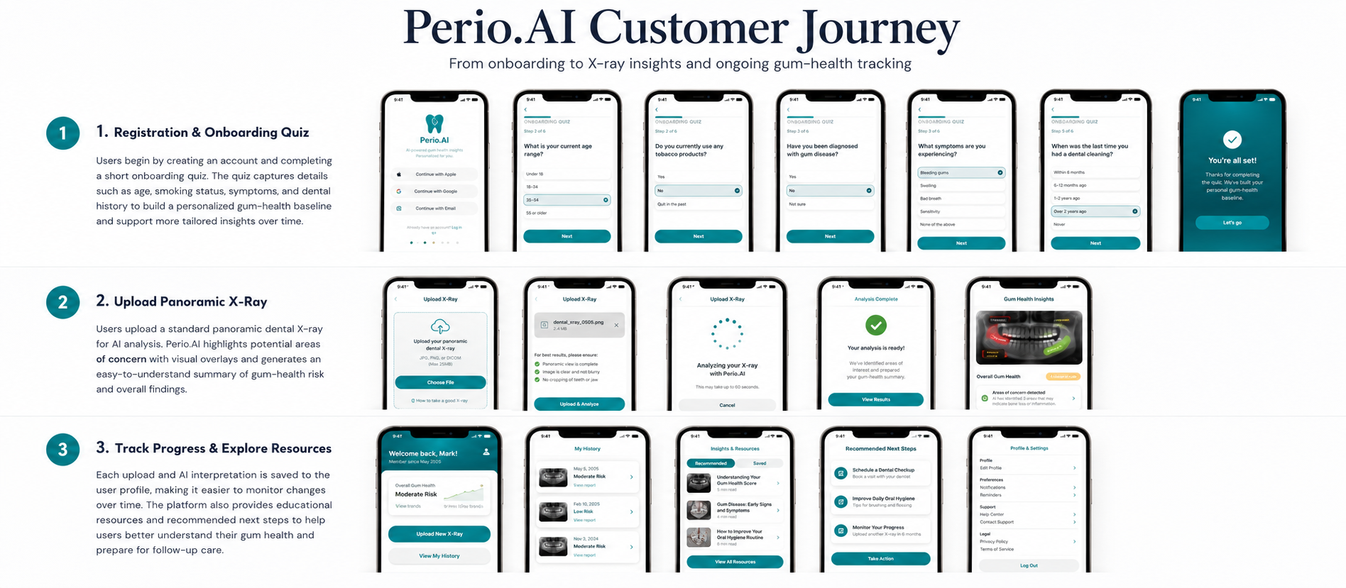
From X-ray to understanding and education — a focused three-step experience designed around clarity, not clinical complexity.
06
Why AI, and model selection
If we train deep learning models on large-scale annotated dental X-rays to a 70%+ confidence level, the system should detect early-stage gum disease with more accuracy, consistency, and explainability than relying on a clinician's read alone.
That called for a dual-model approach rather than a single classifier:
YOLOv8
Object detection — identifies and localises periodontal indicators such as bone loss and structural defects on the X-ray.
U-Net
Segmentation — precisely outlines gum and bone structures so the system can actually measure what YOLOv8 flags.
One visual layer, clarity for both sides. Perio ai translates complex radiographic signals into an accessible report without making treatment decisions itself:
See what matters
Visual overlays highlight the relevant areas of the X-ray.
Understand severity
Plain-language indicators communicate the level of concern.
Start a better conversation
A shared visual reference supports the patient–dentist discussion.
07
Data strategy
Data collection starts at onboarding — an intake quiz capturing age, smoking status, and past gum issues builds a dental health profile for each user, layered on top of the X-ray itself. Every upload and interpretation is recorded to support longitudinal tracking of a patient's gum health over time, not just a single-visit snapshot.
YOLO + U-Net
landmark detection
calculation
confidence-scored result
We developed a data strategy that balanced rapid MVP delivery with a path toward clinical reliability. We used public panoramic X-ray datasets to validate the ML pipeline, while designing a controlled annotation process for tooth masks, CEJ boundaries, bone levels, and root length.
We decomposed the model into measurable stages—image validation, tooth segmentation, landmark detection, bone-loss estimation, severity classification, and confidence filtering—so that performance and failure points could be evaluated independently. The strategy also introduced clinician-reviewed labels, model and dataset versioning, confidence calibration, subgroup testing, and a controlled retraining loop.
This allowed us to demonstrate technical feasibility without presenting the proof of concept as a clinically validated diagnostic system.
08
AI model validation
Designed as a trusted second-opinion layer — Perio ai supports the patient–clinician relationship, it doesn't replace it. Three principles kept that boundary explicit:
Explainable by design
Overlays, severity indicators, and confidence scores make the output visible and interpretable, not a black-box score.
Clinician in the loop
Findings are framed for professional review, not autonomous diagnosis or treatment.
Clear boundaries
Plain language and careful guardrails communicate what the system can — and can't — conclude.
Built from real conversations, not assumptions:
09
User stories & acceptance criteria
Three core screens make up the current feature set:
- 01
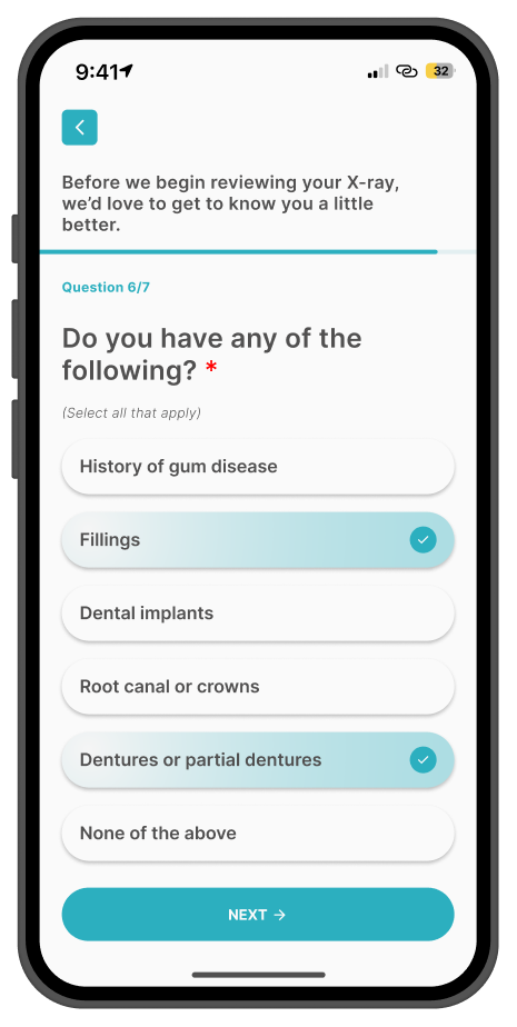
Collects age, smoking status, and past gum issues to build a dental health profile.
- 02
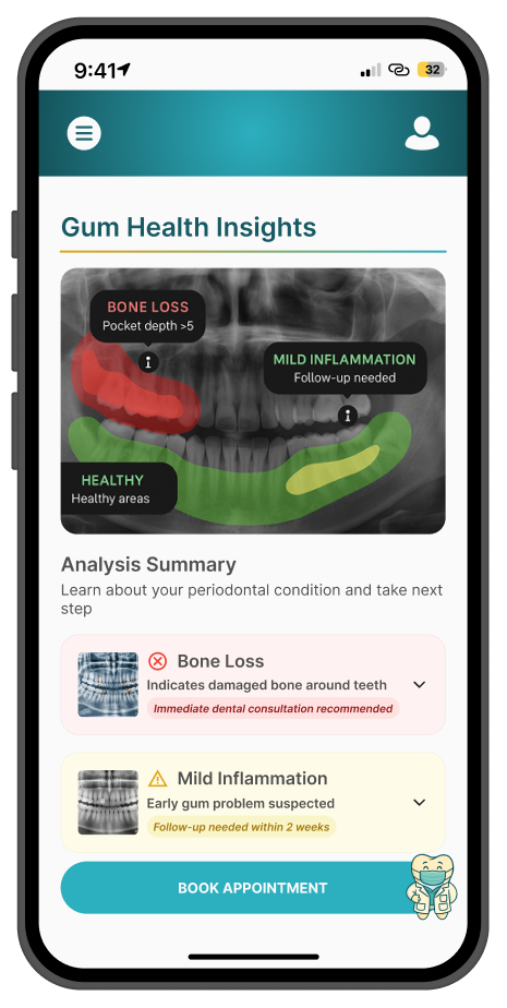
User uploads a U.S.-standard panoramic X-ray; AI generates a heatmap of the affected areas and an overall gum health score.
- 03
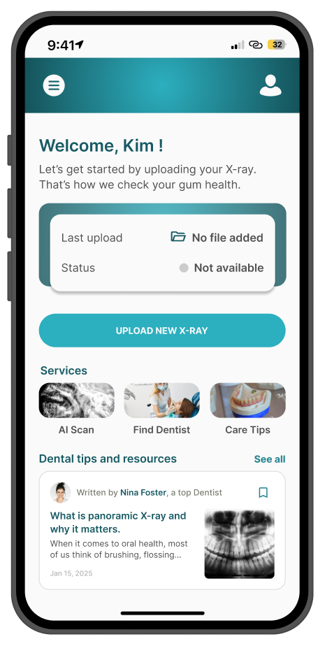
Tracks uploads and interpretations over time, and recommends educational resources and accessible providers.
10
System architecture
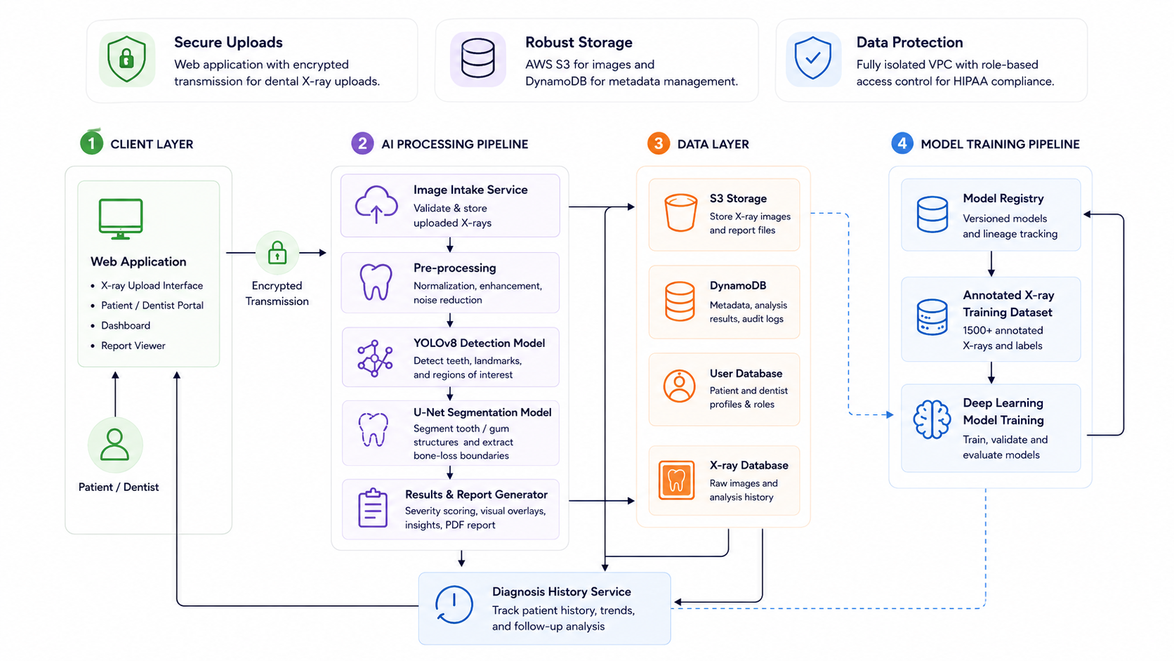
11
Lessons learned
Better-labelled data mattered more than more data.
We initially assumed that improving the model would mainly require a larger volume of panoramic X-rays. However, the annotation exercise revealed that dataset size alone was not enough.
Blur, overlapping anatomy, restorations and missing teeth often made the CEJ and periodontal bone level difficult to identify. Early annotation samples also differed in tooth numbering and landmark representation. Training on inconsistent labels would have taught the model unreliable patterns, regardless of dataset size.
This shifted our priority from acquiring more images to defining better ground truth: a shared annotation protocol, consistent anatomical definitions, an "ungradable" label for unclear regions, and dental-expert review of ambiguous cases.
In healthcare AI, label quality and clinical consistency are more valuable than data volume alone.